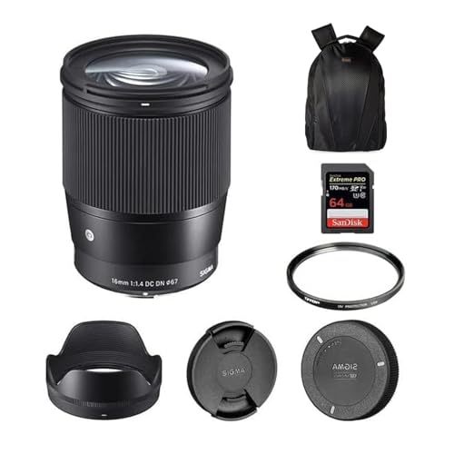
To achieve optimal results with your imaging setup, ensure proper alignment of the optical components. Begin by securely attaching your imaging device to the eyepiece tube or the designated port. This initial step is crucial for capturing clear and focused images.
Next, adjust the illumination settings carefully. Proper lighting enhances contrast and visibility of the specimens. Experiment with different light intensities and angles to find the settings that best highlight details in your samples.
Focusing on your specimens can be achieved by utilizing both coarse and fine focus knobs. Start with coarse adjustments to locate the area of interest, then switch to fine adjustments for precise clarity. Familiarize yourself with the depth of field, which can significantly impact the sharpness of your images.
Utilize imaging software effectively for capturing and analyzing images. Most applications provide features like brightness, contrast adjustments, and measurement tools. Take advantage of these to enhance your data analysis and presentation.
Finally, maintain your equipment regularly. Cleaning the optical surfaces and ensuring the device is free of dust will prolong the lifespan and performance of your system. Make this a part of your routine to avoid common pitfalls.
Using Amscope’s Objective Attachment
Insert the adapter tube into the optical path of your instrument with a secure fit. Align it properly to ensure optimal light transmission. Adjust the height of the apparatus if necessary to achieve a comfortable viewing angle.
Connect the imaging component to your computer or mobile device using the provided USB cable. Ensure that your device recognizes the attachment for immediate access. Launch the appropriate software to facilitate image capture and live feed.
Choose the right optical multiplier based on your sample’s characteristics. A higher magnification can reveal intricate details, while a lower one can offer a broader overview of the specimen. I prefer beginning with a lower optical setting for initial observations.
Fine-tune the focus for clarity. Gradual adjustments will yield the sharpest images; avoid abrupt changes. Utilize the fine focus knob for precise control, ensuring the specimen remains in clear view.
During analysis, I often toggle between various illumination settings. Adequate lighting enhances contrast, highlighting specific features within the specimen. Adjust brightness levels as needed for optimal visibility.
For documentation, capture images or record video through the software interface. Save files with descriptive names for easy retrieval later. Utilize annotations within the program to mark key details relevant to your findings.
Choosing the Right Amscope Microscope Lens
Select a lens based on your specific objectives and subject matter. Consider factors such as magnification levels, resolution requirements, and compatibility with your existing equipment.
Magnification Requirements
- Identify the desired magnification range. For biological samples, a 10x to 40x may suffice.
- For detailed cellular structures or fine details, consider lenses offering 100x or higher magnification.
Resolution and Image Clarity
- Choose high-quality optics for better image clarity and reduced distortion.
- Look for lenses designed with anti-reflective coatings to enhance light transmission.
Also, assess the compatibility with your current imaging setup, ensuring lens dimensions align with the camera sensor size to avoid cropping or other image issues. Always consult specifications before making a final decision.
Setting up the microscope for camera attachment
First, ensure the light source is adequately positioned for optimal illumination. Adjust the brightness to suit the specimen you will be examining to avoid glare or insufficient light.
Securing the Camera
Align the imaging device with the ocular tube. This step involves removing the eyepiece from the tube if necessary and gently inserting the camera adapter. Ensure a snug fit to avoid wobbling during observation.
Focusing for Clarity
Once attached, slowly bring the specimen into focus using the fine focus knob. Avoid using coarse adjustments while the camera is mounted to prevent damage. Utilize the zoom function if available to enhance details.
It’s crucial to calibrate the settings within your software, enabling the capture of high-resolution images. Adjust exposure and white balance according to specimen characteristics.
- Check all connections to prevent disconnections.
- Focus on the specimen before beginning any digital work.
- Regularly clean the lenses to ensure clarity.
After setup, perform a test capture to assess image quality and make any necessary adjustments to settings or positioning.
Connecting the Camera to Your Computer or Monitor
First, ensure the device is powered on and all cables are properly connected. I recommend using a USB cable to link the imaging device to the computer for straightforward setup. The software provided with the device should assist in recognizing the camera upon connection.
Next, install the software if it’s not already on your machine. This typically includes drivers and an interface for viewing captured images. After installation, reboot your computer to complete the setup process.
Once the computer restarts, open the imaging software. Select the connected unit from the list of available devices. Sometimes, toggling between different display modes or resolutions may be necessary to achieve the best quality image on your monitor.
If using an external display, make sure to connect it via HDMI or VGA, depending on your setup. Adjust the display settings accordingly to ensure clarity and proper resolution. Check settings for aspect ratio and scaling if the image appears distorted.
Regularly update the software to benefit from enhancements and fixes. Monitor performance for any lag or connection issues, addressing them promptly by checking cable integrity or software updates. Following these steps allows for optimal function and image quality.
Adjusting the Focus for Optimal Image Clarity
For achieving the best clarity, it’s crucial to fine-tune the focus meticulously. I recommend starting with the lowest magnification objective. This creates a wider field of view, making it easier to locate your specimen.
Step-by-Step Focusing Process
Firstly, position the specimen directly beneath the objective, ensuring it’s illuminated adequately. Slowly, raise the stage until the specimen is close to the lens. Gradually decrease the distance by turning the coarse focus knob. As your image begins to come into view, switch to the fine focus knob for precision adjustments. This will help eliminate any blurriness.
Tips for Maintaining Focus
To maintain optimal clarity, avoid touching the stage or specimen during observation. Any vibrations can lead to loss of focus. Additionally, if switching to a higher magnification, recheck the image and repeat the fine focusing process. Take note of your settings for future reference to streamline the adjustment process later on.
Setting the Appropriate Exposure and Brightness
Adjust exposure settings through the camera software interface. Lowering the exposure time can prevent overexposure when viewing brighter specimens. Conversely, increasing it may be necessary for darker samples to ensure adequate visibility.
Monitor the brightness through real-time feedback on the display. The histogram feature can significantly aid in visibly analyzing the light distribution in the captured images, allowing for precise adjustments to achieve balanced exposure.
Utilizing a neutral density filter can also enhance your experience by reducing light intensity without altering the color balance. This tool is particularly effective for extremely bright environments, ensuring that details remain distinguishable.
Testing different settings is key. Capture a series of images while varying exposure and brightness levels. This practice will help identify the optimal configuration for consistent, high-quality results based on the specimen and lighting conditions.
Regularly calibrate your equipment, as consistent adjustments may be necessary due to changes in ambient lighting during usage. Maintaining a log of preferred settings can save time during future sessions.
Utilizing Software for Capturing Images and Videos
For optimal results with your imaging setup, select software that aligns with your objectives. I recommend starting with options like Amscope’s proprietary software or third-party applications that support camera integration.
Key Features to Look For
- Support for live image preview to assist with adjustments.
- Capability to capture single images or continuous video streams.
- Image processing tools like brightness, contrast, and color adjustments.
- Annotation tools for marking specific areas of interest directly on images.
- Export options for saving files in various formats (JPEG, PNG, AVI).
Steps to Capture Visuals
- Launch the selected software and connect your device following the manufacturer’s instructions.
- Adjust the settings to match your imaging needs, focusing on resolution and frame rate.
- Utilize the live preview feature to frame your subject correctly, ensuring clarity.
- Use the capture button to take still images or initiate video recording as required.
- Save captured visuals to your designated storage location, ensuring to label files for easy retrieval.
Experiment with different settings and features available in the software to enhance your workflow and results. Regularly check for updates to ensure compatibility and access to new tools and features.
Calibrating the Camera for Accurate Measurements
To ensure precise measurements, calibrate the equipment with a stage micrometer. I position the micrometer slide under the optical system, capturing an image at the desired magnification. It’s crucial to note the scale on the micrometer, typically in micrometers (µm) or millimeters (mm).
Next, measure the distance between defined increments on the micrometer image. I focus on two reference points and use image processing software to determine the pixel distance between them. This step helps me establish a conversion factor.
After calculating the pixel-to-length ratio, I input this data into the measurement settings of the imaging software. This allows subsequent measurements to reflect real-world dimensions accurately. I recommend double-checking these settings before proceeding with further imaging tasks.
Finally, perform test measurements on known objects to confirm calibration accuracy. If discrepancies arise, recalibrate as necessary. Consistency in the calibration process enhances reliability in data collection and analysis.
Exploring Different Magnification Options
To achieve desired results, selecting the right level of enlargement is key. Here’s what I consider when exploring available options:
Standard Magnification Levels
Start with the standard objectives, typically ranging from 4x to 100x. Each has its unique purpose:
- 4x: Ideal for scanning larger samples quickly.
- 10x: Great for detailed observation of larger structures.
- 40x: Suitable for examining cell morphology and tissue architecture.
- 100x: Perfect for studying microscopic organisms and intricate details.
Digital Zoom and Software Enhancement
Utilizing digital zoom features in conjunction with optical magnification can enhance clarity:
- Digital zoom can be adjusted within software settings, allowing for further enlargement without significant loss of detail.
- Ensure to balance optical and digital magnification to avoid pixelation.
- Test various combinations of optical and digital settings to find the perfect blend for your specimen.
Experimenting with these different options will help refine my approach and enhance the quality of observations and images captured. Adjusting both optical and digital settings allows for a tailored experience focused on specific requirements of the subject being studied.
Using Illumination Techniques for Enhanced Observation
Adjusting light sources dramatically refines visibility of specimens under scrutiny. In my experience, employing transmitted illumination is ideal for transparent samples, allowing me to see the internal structures clearly. Position the light source beneath the stage and manipulate the diaphragm to regulate brightness.
For opaque materials, reflected illumination works wonders. A directed light beam shines at an angle, revealing surface details. Experimenting with light intensity helps in discerning intricate features without overwhelming the image.
Polarized light enhances contrast significantly, particularly for crystalline substances. By incorporating a polarizing filter, I can highlight specific attributes, making analyses more precise.
Darkfield illumination serves as a remarkable alternative, illuminating specimens against a dark background. This method uncovers the edges and outlines of samples, beneficial for viewing less pigmented microorganisms.
Furthermore, adjusting the angle and intensity of each light source is critical. Small tweaks often yield substantial improvements in clarity and detail, transforming the overall observation. Always assess the specimen under various lighting conditions for the best results.
Using multiple techniques in combination can provide comprehensive insights. For example, a base of brightfield illumination combined with darkfield can enhance underlining details while emphasizing surface characteristics.
Integrating Additional Filters for Better Visibility
Integrating filters enhances clarity by minimizing glare and improving contrast. I recommend a polarizing filter to reduce reflections from shiny surfaces, which can obscure critical details. Ensure it is properly aligned with the optical path for maximum effectiveness.
Color Filters
Color filters can significantly improve observation of certain specimens. For example, a green filter enhances visibility for chlorophyll-rich samples, while a yellow filter helps to highlight details in thin specimens. Selecting the right color filter tailored to your observations can reveal structures that may otherwise remain hidden.
Neutral Density Filters
Neutral density filters are useful for managing brightness without changing color balance. They are particularly beneficial when working with high-intensity light sources. By reducing light intensity evenly across the spectra, these filters prevent the saturation of images and allow for better detail capture.
Saving and Organizing Captured Images Efficiently
To manage images captured through the optical system, I recommend creating a well-structured folder hierarchy on your computer. Organize files by date, subject, or specimen type for easy retrieval later. For instance, I create a main folder labeled ‘Microscopy Images’, with subfolders for each project or specimen type.
File Naming Conventions
Implement descriptive file naming conventions that include relevant details such as specimen name, date, and magnification level. For example, naming a file as ‘Onion_Cell_2023_10_03_40x.jpg’ allows for quick identification. I also suggest avoiding spaces or special characters in file names, using underscores or dashes instead.
Image Backup Strategies
Regularly back up images to cloud storage or external drives. I utilize services like Google Drive or Dropbox for secure off-site storage. Additionally, maintain a secondary backup on an external hard drive to safeguard against data loss. This practice ensures that valuable specimens are preserved even in case of system failures or accidental deletions.
Adjusting Settings for Specific Specimen Types
For optimal results with various specimen types, I adjust multiple parameters. For biological samples like cells or tissues, setting the illumination to brightfield is ideal. For more transparent specimens such as live organisms, I often switch to phase contrast, enhancing visibility.
For mineral samples, a lower light intensity works well, reducing glare and improving texture observation. I frequently employ polarized light, which can reveal intricate crystal structures.
When dealing with stains or colored slides, balancing contrast and brightness becomes a priority to prevent washout. Below is a summary of recommended settings for various sample types:
| Specimen Type | Illumination Technique | Brightness Setting | Contrast Level |
|---|---|---|---|
| Biological Tissues | Brightfield | High | Medium |
| Live Cells | Phase Contrast | Medium | High |
| Minerals | Polarized Light | Low | High |
| Stained Slides | Brightfield | Medium | Adjust as needed |
I also find adjusting the focus finely is more valuable with intricate specimens. This enables a more detailed examination and clearer images, leading to more accurate results.
Understanding the Importance of Camera Resolution
Choosing a higher resolution for your imaging device directly impacts the clarity and quality of specimens captured. A resolution of 1920×1080 pixels is considered full HD, providing a sharp image suitable for most applications. For more detailed analysis, resolutions above 4K (3840×2160 pixels) allow for finer details to be discerned, which is crucial for research and presentations.
Pixel Count and Detail Representation
Be mindful of pixel density. Higher pixel counts enable more detail, which is particularly vital in fields such as biology and materials science. For example, 12-megapixel devices can capture more intricate features of biological specimens, while 20-megapixel options offer additional fidelity.
| Resolution Type | Maximum Detail | Recommended Use |
|---|---|---|
| Full HD (1920×1080) | Good detail for general use | Basic educational and observational tasks |
| 4K (3840×2160) | Very high detail | Professional research and detailed presentations |
| 8K (7680×4320) | Exceptional detail | Specialized research requiring ultra-high resolution |
Impact on Image Processing
Higher resolution images demand more processing power and storage space. Ensure that your computer or device can handle large files without lag. Effective image processing software often includes features that optimize high-resolution images while maintaining clarity. Regular updates of the software suite can significantly enhance usability and performance.
Maintenance tips for your Amscope microscope camera lens
Regularly inspect the objective and eyepiece for dust or debris. Use a soft, lint-free cloth to gently wipe the surfaces. For stubborn spots, a small amount of lens cleaning solution can be applied. Avoid abrasive materials that could scratch the glass.
Store the optical components in a dust-free environment. Utilize lens caps when the equipment is not in use to prevent contamination.
Check connections occasionally for wear or looseness. Tighten any loose screws or fittings to maintain proper alignment and connection integrity. Consider using a desiccant in storage areas to minimize moisture buildup.
Periodically calibrate settings to ensure precise focus. Ensure that all software is updated to the latest versions to take advantage of enhancements. This practice also helps prevent compatibility issues with your imaging system.
Run routine tests to confirm the quality of captured images. If inconsistencies occur, recalibrate lenses or consult user manuals for troubleshooting advice tailored to specific issues.
Educate yourself about best practices for handling components. Minimal handling reduces the risk of smudges and scratches. Always use a protective case for transportation to avoid impact damage.
Schedule regular maintenance checks with a professional service to keep performance at its peak. This helps identify potential issues before they affect functionality.


